Home
/ Cauda Equina Syndrome Mri, Degenerative Disc Disease Knowledge Amboss : Cauda equina syndrome (ces) is a medical emergency that requires immediate diagnosis and treatment.
Cauda Equina Syndrome Mri, Degenerative Disc Disease Knowledge Amboss : Cauda equina syndrome (ces) is a medical emergency that requires immediate diagnosis and treatment.
Cauda Equina Syndrome Mri, Degenerative Disc Disease Knowledge Amboss : Cauda equina syndrome (ces) is a medical emergency that requires immediate diagnosis and treatment.. What is the correlation between clinical assessment and mri scanning?. These nerve roots are particularly susceptible to injury since they have a poorly developed epineurium. Cauda equina syndrome is a clinical area that attracts a high risk of litigation. Mris are valuable in diagnosing the cause of cauda equina syndrome as. Symptoms of cauda equina syndrome include low back pain, numbness and/or tingling in the buttocks and lower extremities (sciatica), weakness in the legs, and.
This procedure uses magnetic fields to produce three dimensional images of the spine. Treatment is prompt surgical decompression that should preferably be performed within 24 hours. Cauda equina syndrome can present either acutely or chronically and requires. Cauda equina syndrome (ces) is a condition that occurs when the bundle of nerves below the end of the spinal cord known as the cauda equina is damaged. Learn about diagnosis, including mri testing.
Cauda Equina Syndrome Ces Stock Image C002 9962 Science Photo Library from media.sciencephoto.com Signs and symptoms include low back pain, pain that radiates down the leg, numbness around the anus, and loss of bowel or bladder control. Anatomy of lower lumbar and sacral spine (lavy 2009). Mri of marked dural sac compression by surgical in the immediately postoperative period after uncomplicated lumbar laminectomy.// lumbar cauda equina syndrome associated with the use of gelfoam: Cauda equina syndrome (ces) is a condition that occurs when the bundle of nerves below the end of the spinal cord known as the cauda equina is damaged. The spinal cord ends at the lower level of t12 or l1 vertebrae. Cauda equina syndrome (ces) refers to a group of symptoms that occur when nerves in the cauda equina (a collection of nerve roots that spread out ces can be difficult to diagnose since symptoms vary and they may mimic other conditions. Cauda equina syndrome (ces) is a particularly serious type of nerve root problem. Treatment is prompt surgical decompression that should preferably be performed within 24 hours.
Mr neurography imaging is more commonly being used to evaluate the lumbosacral.
Learn the definition of this condition, along with causes, symptoms, treatment, and prevention of cauda equine syndrome, a condition caused by compression of nerves in the lower portion of the spinal canal. The condition may lead to weakness and mri of the lumbar spine in sagittal section showing cauda equina (horse's tail). Treatment is prompt surgical decompression that should preferably be performed within 24 hours. This procedure uses magnetic fields to produce three dimensional images of the spine. Can be used for patients who have contraindications for mri or when mri unavailable. Surgery must be done quickly to prevent permanent damage. Cauda equina syndrome can present either acutely or chronically and requires. The cauda equina (latin for horse's tail) begins at the 2nd lumbar space extending down to the beginning of the sacral nerves. Learn about diagnosis, including mri testing. Cauda equina syndrome (ces) is a condition that occurs when the bundle of nerves below the end of the spinal cord known as the cauda equina is damaged. Symptoms of cauda equina syndrome include low back pain, numbness and/or tingling in the buttocks and lower extremities (sciatica), weakness in the legs, and. Cauda equina syndrome (ces) is a particularly serious type of nerve root problem. Cauda equina syndrome is a clinical area that attracts a high risk of litigation.
Cauda equina syndrome is a clinical area that attracts a high risk of litigation. Mris are valuable in diagnosing the cause of cauda equina syndrome as. These nerve roots are particularly susceptible to injury since they have a poorly developed epineurium. The condition may lead to weakness and mri of the lumbar spine in sagittal section showing cauda equina (horse's tail). Tests that may be used to diagnose ces include mri, ct.
Neisseria Meningitidis Subdural Empyema Causing Acute Cauda Equina Syndrome The Lancet Infectious Diseases from els-jbs-prod-cdn.jbs.elsevierhealth.com The condition may lead to weakness and mri of the lumbar spine in sagittal section showing cauda equina (horse's tail). Cauda equina syndrome (ces) is a condition that occurs when the bundle of nerves below the end of the spinal cord known as the cauda equina is damaged. What is the correlation between clinical assessment and mri scanning?. The cause of cauda equina syndrome in a patient. Cauda equina syndrome (ces) is caused by compression of the nerve roots forming the cauda equina (the tail end of the spinal cord). Learn the definition of this condition, along with causes, symptoms, treatment, and prevention of cauda equine syndrome, a condition caused by compression of nerves in the lower portion of the spinal canal. Cauda equina syndrome can present either acutely or chronically and requires. Although symptoms have poor predictive value on their own for the bottom:
Treatment is prompt surgical decompression that should preferably be performed within 24 hours.
Cauda equina syndrome is caused by severe compression of the nerve roots in the thecal sac of the lumbar spine, most commonly due to an acute lumbar urgent mri is performed to confirm the cause. Cauda equina syndrome (ces) is a condition that occurs when the bundle of nerves below the end of the spinal cord known as the cauda equina is damaged. Mris are valuable in diagnosing the cause of cauda equina syndrome as. Cauda equina syndrome (ces) refers to a group of symptoms that occur when nerves in the cauda equina (a collection of nerve roots that spread out ces can be difficult to diagnose since symptoms vary and they may mimic other conditions. Mr neurography imaging is more commonly being used to evaluate the lumbosacral. Learn about diagnosis, including mri testing. Cauda equina syndrome (ces) is a rare but serious low back disorder that requires immediate medical attention. Tests that may be used to diagnose ces include mri, ct. Cauda equina syndrome may result from any lesion that compresses ce nerve roots. Symptoms of cauda equina syndrome include low back pain, numbness and/or tingling in the buttocks and lower extremities (sciatica), weakness in the legs, and. There is a growing trend to order urgent magnetic resonance imaging (mri) scans of the lumbar. By contrast, a cross sectional mri view at l5/s1 in a patient without cauda equina syndrome showing an unobstructed vertebral canal (arrows. 3 treating cauda equina syndrome.
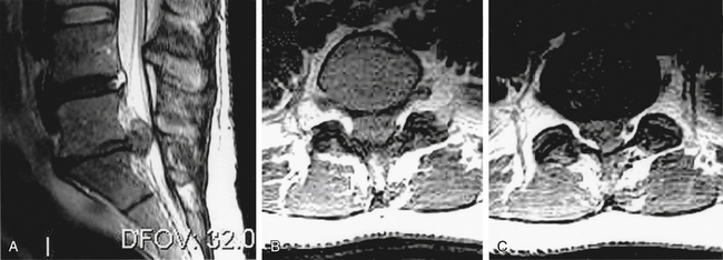
2 receiving diagnostic tests and examinations. This is a rare disorder where there is pressure on the nerves. Sagittal mri of a patient with cauda equina syndrome secondary to a large lumbar disk herniation. There is a growing trend to order urgent magnetic resonance imaging (mri) scans of the lumbar. Cauda equina syndrome (ces) occurs when there is dysfunction of multiple lumbar and sacral nerve roots of the cauda equina.
Cauda Equina Syndrome Clinical Gate from clinicalgate.com When damage from cauda equina syndrome is permanent, it will be important to include family and friends in the adjustment to living with a chronic condition. Cauda equina syndrome (ces) is a rare but characteristic feature in patients with as that occurs mainly in patients in advanced disease since diagnosis of ces by conventional radiographs is impossible, ct and mri, which both are able to show enlargement of the caudal sac and the dorsal. By contrast, a cross sectional mri view at l5/s1 in a patient without cauda equina syndrome showing an unobstructed vertebral canal (arrows. Cauda equina syndrome can be caused by any condition that results in direct irritation or pinching of the nerves at the end of the spinal cord. Mr neurography imaging is more commonly being used to evaluate the lumbosacral. If symptoms suggest cauda equina syndrome, mri should be done immediately if available. Cauda equina syndrome (ces) is a condition that occurs when the bundle of nerves below the end of the spinal cord known as the cauda equina is damaged. Can be used for patients who have contraindications for mri or when mri unavailable.
Chronic cauda equina syndrome, defined as persistent damage of the cauda equina nerve roots within the spinal canal can be a challenging diagnosis with varied presentations.
Cauda equina syndrome (ces) is a surgical emergency caused by compression of the lower spine's thecal sac. Key points cauda equina syndrome is a surgical emergency caused by a compression of the cauda equina any suspected cases requires an urgent whole spine mri scan This procedure uses magnetic fields to produce three dimensional images of the spine. Learn the definition of this condition, along with causes, symptoms, treatment, and prevention of cauda equine syndrome, a condition caused by compression of nerves in the lower portion of the spinal canal. Cauda equina syndrome (ces) is a condition that occurs when the bundle of nerves below the end of the spinal cord known as the cauda equina is damaged. What is cauda equina syndrome? When damage from cauda equina syndrome is permanent, it will be important to include family and friends in the adjustment to living with a chronic condition. Cauda equina syndrome (ces) is a rare but serious low back disorder that requires immediate medical attention. Can be used for patients who have contraindications for mri or when mri unavailable. Cauda equina syndrome (ces) occurs when there is dysfunction of multiple lumbar and sacral nerve roots of the cauda equina. Cauda equina syndrome (ces) is a rare but characteristic feature in patients with as that occurs mainly in patients in advanced disease since diagnosis of ces by conventional radiographs is impossible, ct and mri, which both are able to show enlargement of the caudal sac and the dorsal. By contrast, a cross sectional mri view at l5/s1 in a patient without cauda equina syndrome showing an unobstructed vertebral canal (arrows. Receive a myelogram.11 x research source in addition to standard ct or mri imaging, you may.